What Is a Mammogram? The Complete Guide to Screening, Diagnostic and Results
A patient walked into my office last year holding a letter she had been ignoring for six months. Her primary care doctor had recommended she schedule a mammogram. She kept putting it off , not because she did not care, but because nobody had ever explained what it actually was, what to expect, whether it would hurt, or why the age recommendations she kept reading online were all different.
That six-month delay cost her nothing , her results were normal. But it just as easily could have cost her everything.
This is the guide I wish every woman had before her first mammogram. It covers what a mammogram is and how it works, the difference between a screening and a diagnostic mammogram, what 3D mammography means, exactly what your results say and what to do next, how much a mammogram costs and how to get one free, when you should start based on your age and risk, and what breast cancer actually looks like on a mammogram image. Everything. In plain language. No glossed-over answers.
Mammogram: At a Glance
|
Question
|
Quick Answer
|
|
What is a mammogram?
|
A low-dose X-ray image of the breast. The gold-standard screening tool for breast cancer in the United States.
|
|
What is mammography?
|
The process and technology used to take a mammogram. The procedure; the mammogram is the image.
|
|
Two types
|
Screening mammogram (routine, no symptoms) and diagnostic mammogram (symptom or concern present)
|
|
What is 3D mammography?
|
Digital breast tomosynthesis (DBT) , takes layered images from multiple angles. Finds more cancers, fewer false callbacks.
|
|
Does it hurt?
|
Most women describe mild pressure for a few seconds. Scheduling post-period and taking ibuprofen beforehand helps.
|
|
How long does it take?
|
Screening: 10-20 minutes of imaging. Diagnostic: 30-60+ minutes. Total facility time: 15-45 minutes.
|
|
When should I start?
|
Age 40, per most guidelines. High-risk women may start earlier. The ACS, ACOG, and USPSTF now agree screening should begin in your 40s, though they still differ on how often to screen (see Section on guidelines below).
|
|
How often?
|
Annually ages 40-54; every 1-2 years at 55+. High-risk: annually, often with MRI added.
|
|
Is it covered by insurance?
|
In most cases. Preventive screening mammograms are typically covered under the ACA with no cost-sharing, but coverage can vary by plan and whether the exam is billed as screening or diagnostic , confirm with your insurer.
|
|
How much does it cost?
|
Uninsured: $100-$300. Free programs exist through the CDC (NBCCEDP) for qualifying women.
|
|
Results system
|
BI-RADS 0-6. BI-RADS 1 or 2 = normal/benign. BI-RADS 3 = probably benign, 6-month follow-up. BI-RADS 4 or 5 = biopsy needed.
|
What Is a Mammogram?
A mammogram is a low-dose X-ray image of the breast. The mammography procedure uses a specialized machine to compress and image each breast from multiple angles, producing pictures that allow a trained radiologist to detect cancer, calcifications, cysts, and other abnormalities that cannot be found by physical exam alone.
The word derives from the Latin mamma (breast) and the Greek gramma (image). The procedure is called mammography; the resulting image is the mammogram.
Mammography is the only imaging tool proven in randomized controlled trials to reduce breast cancer mortality at a population level. According to the American Cancer Society, breast cancer mortality in the United States has declined by 44 percent since 1989 , a reduction that has averted approximately 517,900 deaths , driven in large part by widespread mammography screening and improvements in treatment.
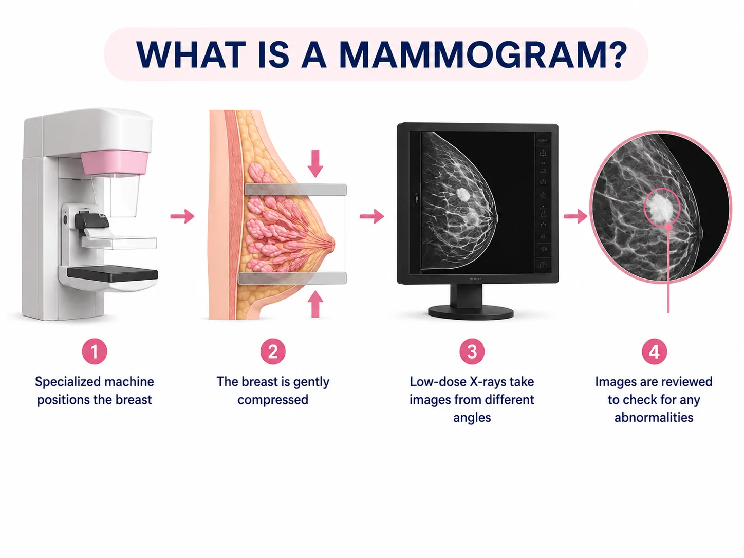
A 2024 RSNA-published analysis of CISNET modeling data found that annual screening starting at age 40 produces a 41.7 percent mortality reduction , significantly higher than biennial screening (25.4% for ages 50-74; 30% for ages 40-74), and with fewer per-mammogram false positives than many alternative scheduling strategies.
Approximately 48 million mammograms are performed in the United States each year. Despite this, studies consistently show that millions of eligible women delay or skip their annual mammogram , often due to anxiety, cost concerns, or simply not understanding what the exam involves.
|
DR. KASHYAP'S TIP
Stage I breast cancer has a five-year survival rate of 99 percent. Stage IV is approximately 32 percent.
|
Types of Mammograms: Screening vs. Diagnostic
Every mammogram is either a screening mammogram or a diagnostic mammogram. These are not interchangeable. They serve different clinical purposes, are billed differently, and result in different procedures at your appointment. Understanding which one you are scheduled for , and why , is important.
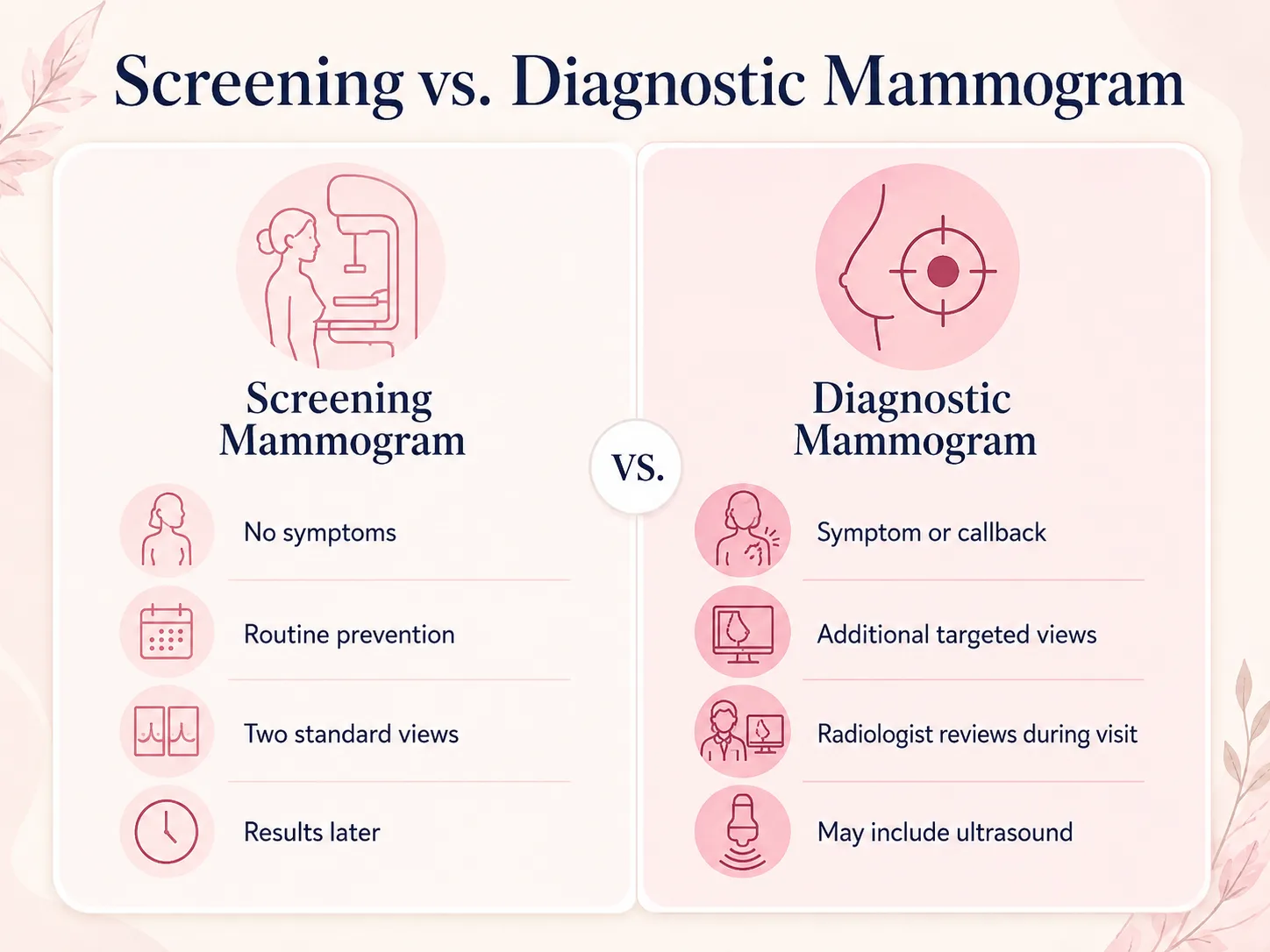
Screening Mammogram: What It Is, What It Finds, and Who Needs It
A screening mammogram is a routine preventive exam performed on women who have no current breast symptoms and no specific concerns. Its purpose is to detect cancer before it causes symptoms , before you or your doctor can feel it, before it has spread, and when treatment is most effective.
Mammogram screening can detect tumors an estimated one to three years, on average, before they become palpable on physical exam. In women under 50, whose tumors may grow faster, this lead time is particularly valuable.
What happens during a screening mammogram
Two standard views are taken of each breast:
- Craniocaudal (CC) , the plate is positioned above and below the breast (top to bottom view)
- Mediolateral Oblique (MLO) , the plate is positioned at an angle from the upper-inner to the lower-outer breast (the most cancer-rich area is captured in this view)
The four images go to a board-certified radiologist who interprets them, often using computer-aided detection (CAD) software to flag potential areas of concern. Results are reported using the BI-RADS scale and sent to your ordering provider, typically within 5-7 days.
What screening mammograms look for
- Calcifications , calcium deposits appearing as white specks. Microcalcifications (tiny, irregular clusters) can indicate early-stage DCIS (ductal carcinoma in situ) or invasive cancer before a lump forms. Macrocalcifications (larger, coarser) are almost always benign.
- Masses or solid lesions , abnormal areas with defined or irregular borders. Shape, margin, and density determine suspicion level.
- Architectural distortion , areas where the normal radial pattern of breast tissue is pulled or distorted, often an early sign of malignanc
- Asymmetry , a difference between the two breasts in density or tissue pattern; new or progressive asymmetry is more significant than stable asymmetry
- Skin thickening or nipple changes , sometimes visible on mammogram even before noticed clinically
Diagnostic Mammogram: When You Need One and What It Involves
A diagnostic mammogram is an expanded, targeted imaging exam ordered when there is a specific concern that requires closer evaluation. It is not a repeat of your screening , it is a different, more comprehensive procedure.
When a diagnostic mammogram is ordered
- A screening mammogram returned a BI-RADS 0 (incomplete , needs more images before the radiologist can give a final read)
- You felt a new lump, thickening, or change in your breast or underarm
- You have nipple discharge , especially spontaneous, one-sided, or bloody discharge
- You noticed skin dimpling, puckering, redness, or an orange-peel texture
- You have a personal history of breast cancer (particularly in the first 3-5 years after treatment, when monitoring is most intensive)
- You have breast implants , standard views do not adequately image implant-augmented breasts; displacement views (Eklund technique) are added
- Your provider wants to follow up a previously identified benign finding at a shorter interval (e.g., a six-month BI-RADS 3 follow-up)
What a diagnostic mammogram involves
Diagnostic mammograms include additional specialized views on top of the standard two-per-breast:
- Spot compression views , a smaller, focused compression paddle pressed directly on the area of concern to spread the tissue and improve visualization
- Magnification views , the X-ray tube is moved away from the breast to enlarge the image and show fine calcification detail
- Additional angles , views from the lateral, medial, tangential, or axillary tail positions depending on where the concern is located
The most important thing I tell patients: being called back for a diagnostic mammogram does not mean you have cancer. The vast majority of callbacks , commonly cited estimates put this at roughly 90 percent , result in benign findings. What it means is that we are doing our job carefully.
Screening vs. Diagnostic Mammogram , Full Comparison
|
|
Screening Mammogram
|
Diagnostic Mammogram
|
|
Purpose
|
Routine cancer detection , no symptoms
|
Investigate a symptom, finding, or abnormal screening result
|
|
Who it is for
|
Women with no breast concerns
|
Women with a lump, discharge, skin change, implants, or a screening callback
|
|
Images taken
|
2 standard views per breast (CC and MLO)
|
Multiple targeted views; spot compression; magnification; additional angles as needed
|
|
Radiologist review
|
After you leave (batch reads)
|
While you are present; preliminary result often given before discharge
|
|
Time for imaging
|
10-20 minutes
|
30-60+ minutes; longer if ultrasound is added
|
|
Results timing
|
Letter or patient portal: 5-7 days
|
Same visit or within 24 hours
|
|
Referral needed
|
Usually not (self-schedule at most centers)
|
Provider order typically required
|
|
Insurance billing
|
Preventive , typically no cost-sharing (ACA), though this can vary by plan
|
Diagnostic , may apply to deductible; confirm with insurer
|
3D Mammography (Digital Breast Tomosynthesis): What It Is and Whether You Need It
Standard mammography produces two-dimensional (2D) flat images of the breast. The limitation: overlapping tissue can create shadows or areas of confusion that either hide a real cancer or look suspicious when nothing is there.
3D mammography , technically called digital breast tomosynthesis (DBT) , addresses this by moving the X-ray tube in an arc around the breast and taking multiple low-dose images from different angles (typically 11-25 projections). These are reconstructed by a computer into a series of thin cross-sectional slices through the breast , like a book of pages rather than a single photograph.
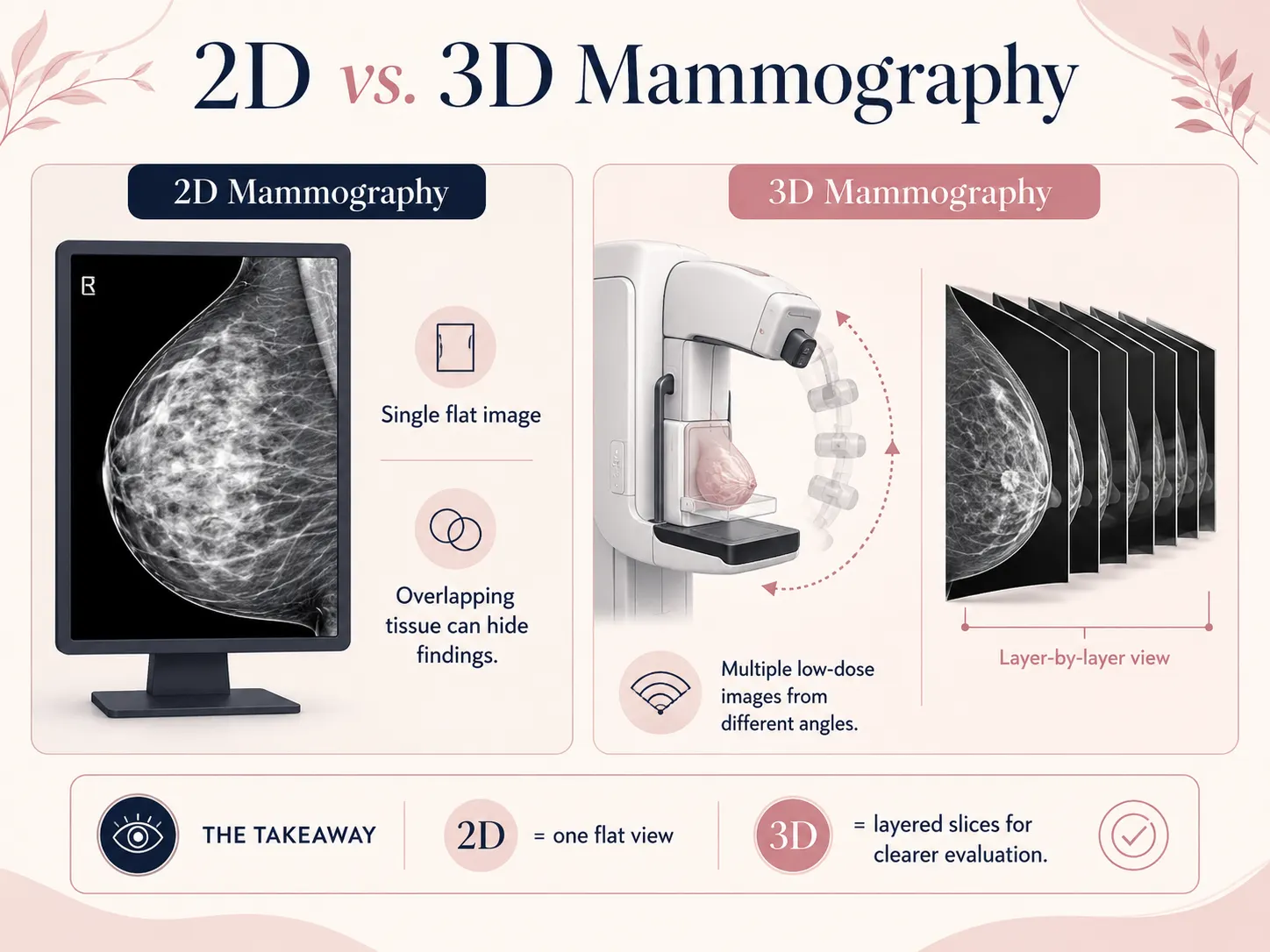
A radiologist scrolls through the slices layer by layer, which means cancers hiding behind overlapping tissue become visible, and areas that looked suspicious in 2D resolve into clearly normal tissue in 3D.
Evidence for 3D mammography
- A major study of more than 450,000 mammograms found 3D mammography detected 41 percent more invasive cancers than 2D alone
- Callback rates are reduced by 15-40 percent with 3D vs. 2D , meaning fewer unnecessary callbacks and the anxiety and cost that come with them
- Benefits are greatest in heterogeneously dense or extremely dense breast tissue , the exact population where 2D is least reliable
- The American Cancer Society, ACR, and Society of Breast Imaging all support 3D mammography as equivalent to or preferred over 2D for breast cancer screening
- Most major mammography centers in the US now offer DBT as the standard modality
3D mammography and radiation
A concern patients often raise: since more images are taken, does 3D mammography expose you to more radiation? The short answer: minimally, and well within safe limits. When 3D is combined with synthetic 2D reconstruction (which generates a traditional-looking 2D image from the 3D data without an additional exposure), total dose is comparable to standard 2D mammography. The average dose is approximately 3.7 mGy , equivalent to about 7 weeks of natural background radiation.
|
QUICK QUESTION , Is 3D the same as a breast MRI?
No. A 3D mammogram still uses low-dose X-rays , just captured from multiple angles. A breast MRI uses magnetic resonance and contrast dye, takes 30-60 minutes, and is significantly more expensive. MRI is more sensitive than mammography but also produces more false positives. It is recommended as supplemental screening for women at high lifetime risk (20%+), not as a routine replacement for mammography in average-risk women. For most women, 3D mammography is the right starting point.
|
How Does a Mammogram Work? The Procedure, Step by Step
A mammogram is performed on a dedicated, FDA-regulated mammography machine. Here is exactly what happens, from the moment you arrive to the moment you leave.
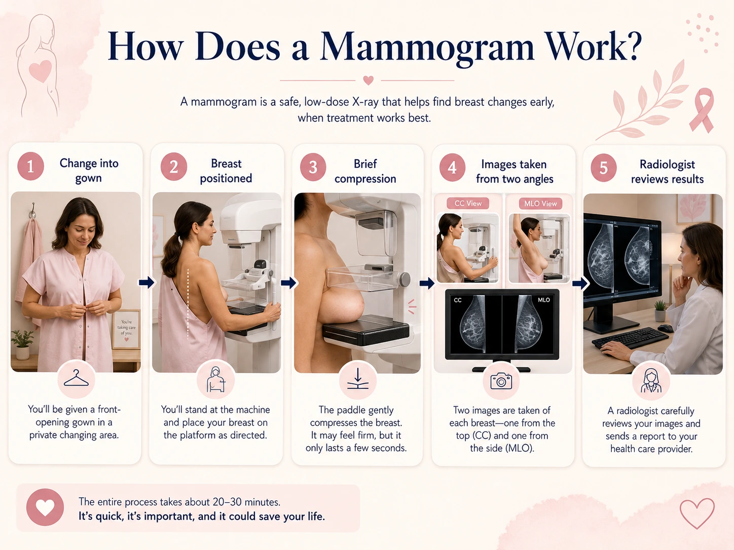
What to Expect During a Mammogram
1. Check-in and gown , You arrive, check in, and are brought to a private changing room. You undress from the waist up and put on a front-opening gown. A female mammography technologist , a certified radiologic technologist with specific mammography training , will be performing your exam.
2. Positioning , The technologist positions you in front of the mammography machine. Your breast is placed on a flat imaging plate called the image receptor. She adjusts your position carefully to ensure the correct amount of tissue is included in the image , this step matters for image quality and is worth taking the extra few seconds to get right.
3. Compression , A second plate , the compression paddle , lowers from above and presses your breast flat between the two plates. This is the step that concerns most women. The compression serves critical purposes: it spreads the tissue evenly so overlapping structures do not hide findings; it reduces motion blur; it reduces radiation dose; and it improves overall image quality. The pressure lasts only a few seconds per image. You can always ask the technologist to pause if you need a moment.
4. Image capture , The X-ray is taken. The compression is released within seconds. For 3D mammography, the tube moves in an arc during acquisition, taking about 4 seconds per view.
5. Repositioning , The technologist repositions your breast and repeats the process from the second angle (MLO view). Then the same is done for the other breast. Four total images for a standard screening; more if diagnostic.
6. Radiologist review , After you leave, a board-certified radiologist reads your images. At a diagnostic mammogram, the radiologist reads while you wait. For screening, results are typically returned within 5-7 business days.
Does a Mammogram Hurt?
This is the most common question I hear from first-time patients: most women experience pressure or mild discomfort, not pain. On a 1-10 scale, most rate their experience a 2-4. A meaningful minority find it uncomfortable enough to wince during compression. A small number find it genuinely painful , usually women with naturally tender breasts, fibrocystic tissue, or those who schedule mid-cycle when breast sensitivity peaks.
What the compression actually feels like: imagine pressing your hand flat on a table and someone laying a book on top. Not pleasant, but not the sharp or stabbing sensation most women fear. The key fact: it lasts 3-7 seconds per image. Four images. Total compression time: under 30 seconds.
How to make a mammogram more comfortable
- Schedule for one week after your period ends , breast tissue is least tender then
- Take 400-600 mg ibuprofen (Advil, Motrin) 30-60 minutes before your appointment (ask your doctor first if you take blood thinners or have kidney issues)
- Tell the technologist if you are especially sensitive , she can adjust compression speed and talk you through each step
- Do not apply lotion, deodorant, or powder to your breast or underarm area that day , they can interfere with image quality and create false findings
- Breathe slowly during compression , holding your breath tensing your muscles can make it feel worse
If pain during mammography has caused you to avoid or delay your exam in the past, please tell your provider. There are strategies , including taking an over-the-counter pain reliever beforehand, scheduling at a less-sensitive point in your cycle, and communicating with the technologist , that genuinely help. Avoiding the exam is never the answer.
How to Prepare for a Mammogram
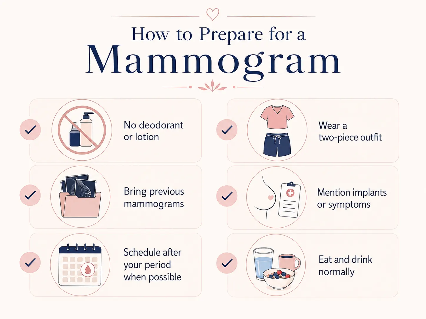
Minimal preparation is required , but getting it right makes a difference in image quality and your comfort.
The day before
- If you have had mammograms at a different facility: request your prior images or imaging records. Most digital images are available on CD. A radiologist comparing your current mammogram to your baseline from two or three years ago is dramatically more accurate than reading a first-time image with no context.
- Gather your breast health history: prior biopsies, surgeries, implants, prior abnormal results. The technologist will ask.
Day of your mammogram
- Do not apply deodorant, antiperspirant, powder, lotion, perfume, or body spray to the breast or underarm area. These products contain particles that appear as white spots on mammogram images , they can look identical to microcalcifications and trigger unnecessary callbacks.
- Wear a two-piece outfit (top and bottom separately) , you will only need to undress from the waist up.
- If you take ibuprofen for discomfort: take it 30-60 minutes before your appointment time, not while you are in the waiting room.
- Avoid scheduling within one week of your period if you have breast tenderness related to your cycle.
- Tell the technologist if you have breast implants , additional displacement views (Eklund views) will be taken to see breast tissue around and behind the implant.
- Tell the technologist about any prior surgeries, lumps, areas of concern, or breast symptoms so she can target the correct areas.
You do not need to fast before a mammogram. Eat and drink normally. There are no medication restrictions unless your provider has specified otherwise.
How Long Does a Mammogram Take?
|
Type
|
Imaging Time
|
Total Facility Time (including check-in)
|
Notes
|
|
Screening mammogram (2D)
|
10-15 minutes
|
20-30 minutes
|
Results in 5-7 days by mail or portal
|
|
Screening mammogram (3D)
|
12-20 minutes
|
20-35 minutes
|
Slight extra time for tomosynthesis acquisition
|
|
Diagnostic mammogram
|
30-60 minutes
|
45-90 minutes
|
Radiologist reviews while you wait; ultrasound may be added
|
|
Diagnostic mammogram with ultrasound
|
45-75 minutes
|
60-120 minutes
|
Allow extra time if you know ultrasound is planned
|
Actual wait times at specific facilities vary considerably. Morning appointments and mid-week slots typically run on schedule; end-of-day Friday appointments at busy hospital imaging centers often run late. It is always worth calling ahead to ask about typical wait times.
Understanding Mammogram Results: The BI-RADS System Explained
Every mammogram report in the United States is structured around the BI-RADS system , the Breast Imaging Reporting and Data System developed by the American College of Radiology. BI-RADS was designed to standardize how radiologists communicate their findings and give every result a corresponding recommended action, so your provider knows exactly what to do next.
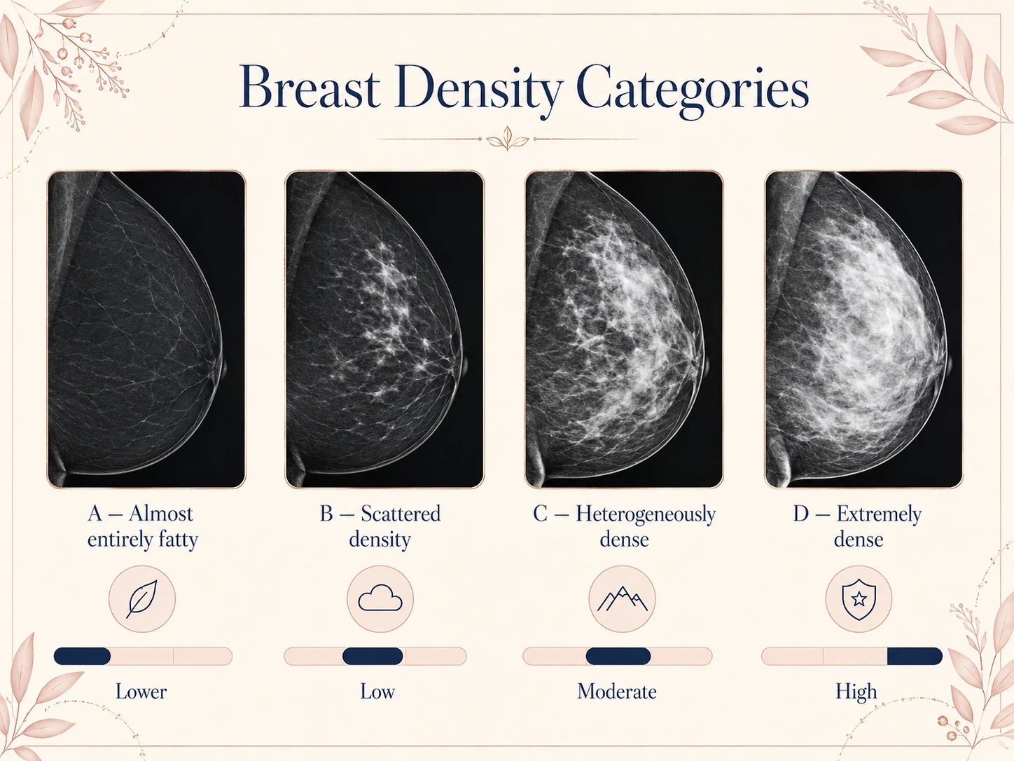
Your report will contain: your BI-RADS category (0-6), a description of findings, and your breast density category (A-D). Always ask for the actual report , not just a verbal summary.
BI-RADS Categories: Full Explanation
|
Category
|
Assessment
|
What It Means
|
Next Step
|
Likelihood of Cancer
|
|
BI-RADS 0
|
Incomplete
|
More imaging needed; the radiologist cannot give a final read from the available images
|
Return for additional views or comparison with prior mammograms
|
Cannot determine yet
|
|
BI-RADS 1
|
Negative
|
Nothing abnormal found; normal mammogram
|
Continue annual screening
|
Essentially zero
|
|
BI-RADS 2
|
Benign
|
A definite finding is present (cyst, calcified fibroadenoma, etc.) but it is clearly benign
|
Continue annual screening
|
Essentially zero
|
|
BI-RADS 3
|
Probably Benign
|
Very likely benign finding (>98% probability not cancer) , but short-term follow-up is recommended to confirm stability
|
Return in 6 months for comparison mammogram
|
Less than 2%
|
|
BI-RADS 4A
|
Low Suspicion
|
Suspicious finding with low probability of malignancy
|
Biopsy recommended
|
2-10%
|
|
BI-RADS 4B
|
Moderate Suspicion
|
Intermediate probability of malignancy
|
Biopsy recommended
|
10-50%
|
|
BI-RADS 4C
|
High Suspicion
|
Moderate concern for malignancy but not classic for cancer
|
Biopsy recommended
|
50-95%
|
|
BI-RADS 5
|
Highly Suggestive
|
Highly suspicious; classic appearance of malignancy
|
Biopsy strongly recommended
|
Greater than 95%
|
|
BI-RADS 6
|
Known Malignancy
|
Biopsy-proven cancer already confirmed; imaging being performed for treatment planning
|
Treatment planning; surgical or oncology referral
|
Confirmed
|
When you receive your results: a BI-RADS 1 or 2 means return to your normal screening schedule. A BI-RADS 3 means follow-up in six months , this is not a biopsy recommendation, just a shorter follow-up. BI-RADS 4 or 5 means a biopsy is recommended, not performed immediately , you will be referred to a surgeon or breast specialist to discuss next steps. Remember: most biopsies come back benign.
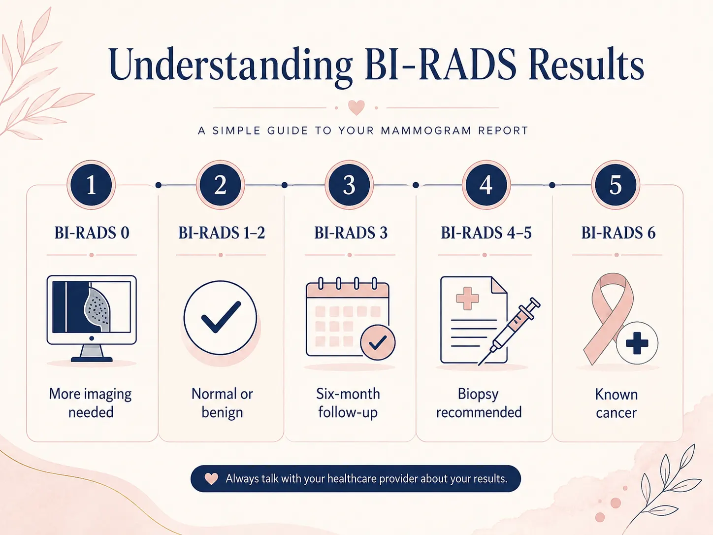
If you receive a BI-RADS 0: call your imaging center. This sometimes happens because prior films were not available for comparison, and showing them resolves the incomplete read without any further imaging.
Your Breast Density Report
Since September 2024, the FDA requires all mammography facilities to notify patients of their breast density in addition to their BI-RADS result. The rule was finalized in March 2023, but enforcement did not begin until September 10, 2024. This was a long-overdue regulatory update , breast density is clinically important and patients deserve to know it.
|
Density Category
|
Description
|
Percentage of Women
|
Cancer Detection Challenge
|
Independent Risk Factor
|
|
A , Almost entirely fatty
|
Very little dense tissue; fatty tissue is dominant
|
About 10%
|
Lowest , fatty tissue appears dark/translucent; cancers appear white and stand out clearly
|
Lowest relative risk
|
|
B , Scattered fibroglandular
|
Mostly fatty with scattered areas of dense tissue
|
About 40%
|
Low , most cancers are still visible
|
Slightly elevated vs. Category A
|
|
C , Heterogeneously dense
|
More dense than fatty; some areas may obscure small cancers
|
About 40%
|
Moderate , cancers can be masked by overlapping dense tissue
|
Moderately elevated; about 1.2x average risk
|
|
D , Extremely dense
|
Almost entirely dense tissue; highest masking risk
|
About 10%
|
High , often described as finding a snowball in a snowstorm
|
Up to 4-6x average lifetime risk vs. Category A
|
Women in Category C or D are considered to have dense breasts. If your report says your breasts are dense, you should discuss whether supplemental screening is appropriate. Most guidelines recommend supplemental ultrasound or MRI for high-density women who also have elevated risk based on other factors. Density alone, in an otherwise average-risk woman, typically results in a discussion , not an automatic MRI order.
|
DR. KASHYAP'S TIP
I have seen women with Category D density who were sent their normal mammogram result and told nothing else. They had no idea their mammogram's ability to find a cancer was significantly reduced. If your report says "extremely dense" or "heterogeneously dense," that is a conversation you need to have , not a footnote to file away. Bring the report to your next appointment and ask: given my density and my overall risk profile, is supplemental imaging appropriate for me?
|
What Does Breast Cancer Look Like on a Mammogram?
This is one of the most-searched mammogram questions online , and one of the most important to answer accurately, because understanding what radiologists look for helps women understand why regular screening matters even when you feel completely well.
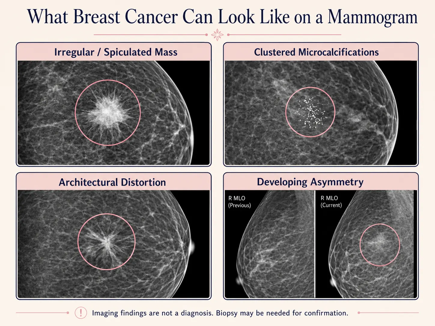
On a mammogram, dense or abnormal tissue appears white and fatty tissue appears dark gray or translucent. Breast cancer most commonly shows up as one or more of these patterns:
1. A mass with irregular or spiculated edges
The most classic appearance of invasive breast cancer on a mammogram is a dense mass with irregular, spiky (spiculated) margins , like a starburst or sea-urchin shape. The spicules represent the cancer invading and pulling on surrounding tissue. In contrast, benign masses (such as fibroadenomas or cysts) typically have smooth, well-defined, round or oval margins. Irregular or microlobulated margins increase radiologist suspicion significantly.
2. Clusters of microcalcifications
Microcalcifications are tiny calcium deposits that appear as small white specks on a mammogram. Groups or clusters of microcalcifications , particularly in a linear or branching pattern (following the path of a duct) , are associated with early breast cancer, especially DCIS (ductal carcinoma in situ). DCIS is a pre-invasive form of breast cancer; finding it on a mammogram through microcalcifications is one of the clearest examples of mammography catching cancer before it becomes invasive. Solitary or scattered round microcalcifications in older women are almost always benign (degenerative changes, vascular calcifications).
3. Architectural distortion
Architectural distortion appears as a pulling or puckering of the normal radial pattern of breast tissue , as though the tissue is being drawn toward a central point. There may be no visible mass. This pattern is associated with invasive lobular carcinoma (which does not always form a distinct lump even clinically) and with radial scars (which are benign but warrant biopsy to confirm).
4. Asymmetry
Radiologists compare the two breasts side by side. A focal asymmetry , a localized area of density present on one breast but not the other , or a developing asymmetry (tissue that appears or increases compared to a prior mammogram) is considered suspicious, particularly when accompanied by other findings.
5. Skin thickening (inflammatory breast cancer)
Inflammatory breast cancer (IBC) is the exception to many mammography rules. It often does not appear as a discrete mass. Instead, it may show as diffuse skin thickening, trabecular (structural) distortion, and increased overall breast density. IBC is rare (1-5% of breast cancers) but aggressive. Redness, warmth, and pitting skin texture clinically , with mammographic skin thickening , together warrant urgent evaluation.
It is important to understand that no mammogram appearance is 100% diagnostic for cancer. That is why BI-RADS 4 and 5 findings result in a biopsy recommendation, not a cancer diagnosis. Only pathology from a biopsy specimen can confirm malignancy.
When Should You Get a Mammogram? Age, Frequency, and Guidelines
This is the topic that generates more confusion than any other in breast health , because the major guidelines organizations in the United States have historically disagreed, and news coverage of their differences has left many women unsure what to do.
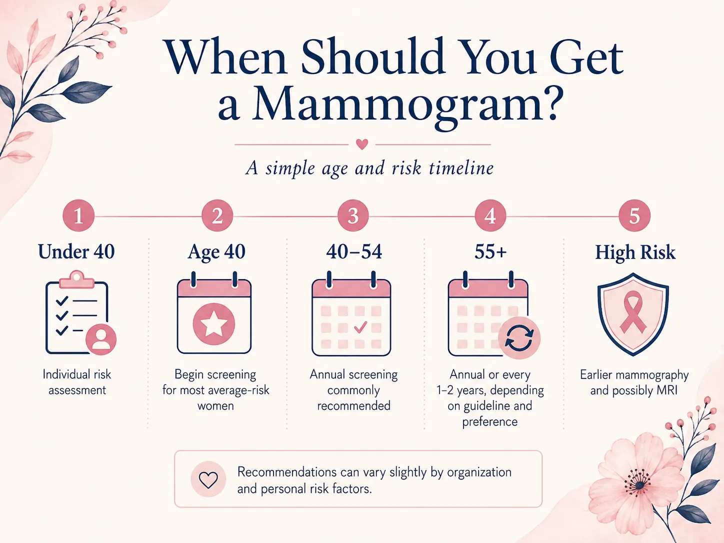
The good news: as of 2024, the major guidelines have converged significantly. The USPSTF updated its recommendation to biennial screening starting at age 40 (previously 50). The ACS recommends annual screening from 45 with the option to start at 40. ACOG recommends offering mammograms annually from 40. All three now agree the decade of the 40s is not to be skipped.
What Age Should You Start?
|
Organization
|
Recommended Start Age
|
Frequency
|
Notes
|
|
American Cancer Society (ACS)
|
45 (option to start at 40)
|
Annual ages 45-54; every 1-2 years at 55+
|
Women 40-44 should have the choice to start; women who want annual starting at 40 should be able to access it
|
|
USPSTF (updated 2024)
|
40
|
Every 2 years
|
Significant update from previous 50; now aligned with other organizations on starting age
|
|
ACOG (American College of OB/GYN)
|
40
|
Annual
|
Supports individual choice; annual preferred
|
|
ACR / Society of Breast Imaging
|
40
|
Annual
|
Annual screening clearly recommended; supports starting earlier in high-risk women
|
|
Dr. Kashyap's practice
|
40
|
Annual
|
Annual from 40; the evidence for annual vs. biennial is debated but I prefer not to give cancer a 12-month head start
|
The debate between annual vs. every-two-years screening comes down to a tradeoff: annual screening catches more cancers at earlier stages but results in more callbacks and false positives. Biennial screening reduces callbacks and overdiagnosis concerns but misses more interval cancers (cancers that develop between screenings). For women with dense breasts, a known mutation, or strong family history, the choice is clearer: annual, full stop.
When to stop: most guidelines recommend continuing screening as long as a woman is in good health and has a life expectancy of at least 10 years. Age alone is not a reason to stop screening for a healthy woman in her 70s.
High-Risk Mammogram Recommendations
Women at higher than average lifetime breast cancer risk have different screening needs. The threshold that triggers supplemental MRI in addition to mammography is a lifetime risk of 20 percent or greater based on a validated risk model (such as Tyrer-Cuzick).
Indications for earlier, more intensive, or supplemental screening:
- Known BRCA1 or BRCA2 mutation , annual mammogram AND annual breast MRI, starting at 25-30 depending on mutation type and family history
- Other hereditary mutations (PALB2, CHEK2, ATM) , enhanced screening; frequency varies by mutation and overall risk model score
- First-degree relative with breast cancer , start screening 10 years earlier than the age of that relative's diagnosis, or at 40, whichever comes first
- Prior chest radiation before age 30 (lymphoma, childhood cancers) , annual mammogram AND annual MRI starting 8-10 years after radiation, or at age 25
- Prior biopsy showing ADH, ALH, or LCIS , increased surveillance; medication to reduce risk may be discussed
- Lifetime risk 20% or greater on a formal risk model , annual mammogram AND annual breast MRI
Finding a mammogram near you
To find an accredited mammography facility near you:
- Use the FDA MQSA Facility Search tool at fda.gov/radiation-emitting-products/mqsa-insights/mammography-facility-search , every facility that accepts Medicare must be FDA-MQSA certified and is listed
- Ask your OB-GYN, primary care provider, or gynecologist for an order or self-referral , most screening mammograms do not require a referral and can be self-scheduled
- Call your insurance company's member services line and ask which mammography facilities are in-network
- Contact your local hospital system's women's imaging or breast center , most accept self-referrals for screening
|
DR. KASHYAP'S TIP
If you are in the Henderson or Las Vegas Valley area: Galleria Women's Health provides comprehensive breast health consultations , risk assessment using validated models, and coordination of your mammogram and imaging referrals with an accredited facility (we do not perform mammography in our office). You should not be navigating any of this alone. Our office can help you understand your full picture, not just your mammogram result.
|
Common Mammogram Myths , Debunked
Myth 1: "I can feel a lump, so I would know if I had cancer."
False. Mammography detects tumors 1-3 years before they become palpable. Small, early-stage tumors , particularly DCIS , often have no symptoms whatsoever. The women whose cancers are caught at Stage I are almost always the ones who had mammograms, not the ones who found a lump.
Myth 2: "Mammograms cause cancer from the radiation."
The radiation dose from a mammogram is extremely low , approximately 0.4 millisieverts (mSv) per study, equivalent to about 7 weeks of natural background radiation. For context, a transatlantic flight exposes you to approximately 0.1 mSv. The consensus of every major radiology and oncology organization in the world: the benefit of mammography screening far outweighs any theoretical radiation risk. This myth keeps women from screening and costs lives.
Myth 3: "My breasts are too small / too large for a mammogram."
False. Mammography can be performed on virtually any breast size. The technologist will work with you to get adequate positioning regardless of breast size. Women with very small breasts may find positioning slightly more technically challenging, but it is not a barrier to getting a mammogram.
Myth 4: "I had a normal mammogram last year, so I do not need one this year."
A normal mammogram result tells you about last year, not this year. Breast cancer can develop quickly , interval cancers (diagnosed between scheduled screenings) account for 20-30% of cancers in screened women. Annual mammography is annual because biology does not take a year off.
Myth 5: "Mammograms are only for women with family history."
Approximately 85 percent of women who develop breast cancer have no family history of the disease. The single most common risk factor for breast cancer is being female and aging. Every woman is at risk. Mammography screening guidelines apply to all women, not just those with family history.
Myth 6: "If I have dense breasts, my mammogram is useless."
Not accurate. A mammogram in a woman with dense breasts is still clinically useful , it finds many cancers, just with reduced sensitivity vs. a fatty-breast mammogram. 3D mammography significantly improves cancer detection in dense tissue. The appropriate response to a dense breast report is to discuss your overall risk picture and whether supplemental imaging is warranted, not to stop screening.
The Bottom Line
A mammogram is the most proven tool we have for catching breast cancer before it kills. It is not optional. It is not just for women with family history. It is not something you wait until you feel something to schedule.
Breast cancer mortality in the United States has dropped 44 percent since 1989. That number was built on mammography, research, and women who showed up for their appointments.
If you are 40 and have not had a mammogram this year, that is the action item , stay current with your screening schedule.
If you have a history, a mutation, a prior biopsy, or density concerns , and no one has ever done a formal risk assessment with you , that is a conversation worth having with a breast health specialist.
[Stay current with your recommended screening schedule, and reach out to Galleria Women's Health in Henderson, NV for a breast health consultation or risk assessment if you have questions about your personal risk, breast density, or a mammogram result]
Key Terms: Mammogram Glossary
- Plain-language definitions for every term you will encounter in your mammogram report, imaging results, and this guide:
- Mammogram , The low-dose X-ray image of the breast itself. Also used informally to refer to the entire exam.
- Mammography , The imaging procedure and technology used to take a mammogram. The process; the mammogram is the result.
- Screening mammogram , A routine preventive mammogram for women with no breast symptoms. Typically four total images (two per breast). Results in 5-7 days.
- Diagnostic mammogram , A targeted mammogram for women with a symptom, concern, or prior abnormal finding. More images; radiologist reviews while you wait.
- Digital breast tomosynthesis (DBT) , 3D mammography. The X-ray tube moves in an arc and takes multiple images that are reconstructed into layered slices. Finds more cancers; reduces callbacks. Now the standard modality at most US centers.
- BI-RADS , Breast Imaging Reporting and Data System. The 0-6 scale used by radiologists to categorize mammogram findings and recommend next steps. Standardized and required by the FDA.
- BI-RADS 0 , Incomplete , additional imaging needed before the radiologist can give a final assessment.
- BI-RADS 1 , Negative , nothing abnormal found. Resume annual screening.
- BI-RADS 2 , Benign finding , a definite finding is present and confirmed benign. Resume annual screening.
- BI-RADS 3 , Probably benign , >98% likely not cancer; follow up in 6 months to confirm stability.
- BI-RADS 4 (4A/4B/4C) , Suspicious , biopsy recommended. 4A: low suspicion (2-10%). 4B: intermediate (10-50%). 4C: high suspicion (50-95%).
- BI-RADS 5 , Highly suggestive of malignancy , >95% probability of cancer. Biopsy strongly recommended.
- BI-RADS 6 , Known biopsy-proven malignancy. Imaging performed for treatment planning.
- Microcalcifications , Tiny calcium deposits (less than 0.5 mm) visible as small white specks. Clusters in linear or branching patterns can indicate DCIS or invasive cancer.
- Macrocalcifications , Larger, coarser calcium deposits. Almost always benign (degenerative changes, old injury, arterial calcifications).
- Architectural distortion , Pulling or puckering of the normal tissue pattern toward a central point, without a visible mass. Associated with invasive lobular carcinoma and radial scars.
- Asymmetry , A difference in density or tissue distribution between the two breasts. Developing or focal asymmetry is more significant than global or stable asymmetry.
- Breast density , The ratio of fibroglandular tissue to fatty tissue on a mammogram. Reported in four categories (A-D). Federal disclosure to patients has been required since enforcement began on September 10, 2024 (the rule was finalized in March 2023). Higher density masks cancer and independently raises risk.
- Callback / recall , Notification that additional imaging is needed after a screening mammogram. Approximately 10-14% of women are called back; fewer than 1 in 10 callbacks result in cancer.
- Spot compression , A smaller compression paddle pressed directly on a specific area of the breast to spread overlapping tissue and improve visualization of that region.
- MLO view (Mediolateral Oblique) , One of the two standard mammogram angles. Captures the breast from upper-inner to lower-outer; the axillary tail (where most cancers occur) is included.
- CC view (Craniocaudal) , The second standard mammogram angle. Top-to-bottom view of the breast.
- DCIS , Ductal carcinoma in situ. Pre-invasive cancer confined to the milk ducts. Often detected by microcalcifications on mammogram before any symptom develops. Highly treatable.
- False positive , A mammogram result that looks suspicious but turns out to be benign after further testing. Common (~10-14% of screens) and anxiety-producing but rarely indicates cancer.
- False negative , A mammogram that misses a cancer that is actually present. Published estimates commonly range from 10-15% of cases, though rates vary by study and breast density. Most common in dense breast tissue. Reason supplemental imaging is recommended for high-risk women.
- Tyrer-Cuzick model , A validated mathematical model that calculates lifetime breast cancer risk by incorporating family history, density, prior biopsy results, hormonal factors, and other variables. Risk of 20%+ triggers supplemental MRI recommendation.
- NBCCEDP , National Breast and Cervical Cancer Early Detection Program. CDC-funded program providing free or low-cost mammograms to qualifying low-income, uninsured, and underinsured women in every US state.
- MQSA , Mammography Quality Standards Act. Federal law requiring all US mammography facilities to be FDA-certified and meet specific quality standards for equipment, personnel, and image quality.
- Gadolinium , The contrast dye used in breast MRI (not mammography). Injected intravenously; highlights areas of increased blood flow associated with tumors. Not used in standard mammography.
Sources and References
1. American Cancer Society. Breast Cancer Statistics, 2024 (Giaquinto et al.). CA: A Cancer Journal for Clinicians. October 2024.
2. Monticciolo DL, Hendrick RE, Helvie MA. Outcomes of Breast Cancer Screening Strategies Based on CISNET Estimates. Radiology, February 2024. (41.7% mortality reduction with annual screening ages 40-79.)
3. U.S. Preventive Services Task Force. “Breast Cancer: Screening.” Updated April 30, 2024. Recommendation: biennial mammography beginning at age 40 for all women at average risk.
4. American Cancer Society. ACS Breast Cancer Screening Guidelines.
5. Siu AL; U.S. Preventive Services Task Force. “Screening for Breast Cancer: U.S. Preventive Services Task Force Recommendation Statement.” Annals of Internal Medicine, 2024.
6. Lee SJ et al. “The Screening Value of Mammography for Breast Cancer: An Overview of 28 Systematic Reviews with Evidence Mapping.” Journal of Cancer Research and Clinical Oncology, 2025. PMC11885354.
7. FDA. Mammography Quality Standards Act (MQSA) and MQSA Program. Updated 2024.
8. Centers for Disease Control and Prevention. National Breast and Cervical Cancer Early Detection Program (NBCCEDP).
9. ACR BI-RADS Atlas, 5th Edition. American College of Radiology, 2013.
10. Conant EF et al. “Association of Digital Breast Tomosynthesis vs Digital Mammography with Cancer Detection and Recall Rates by Age and Breast Density.” JAMA Oncology, 2019.
11. National Cancer Institute. Survival Rates and Prognosis for Breast Cancer (SEER Program data).
12. Friedewald SM, Rafferty EA, Rose SL, et al. Breast Cancer Screening Using Tomosynthesis in Combination With Digital Mammography. JAMA. 2014;311(24):2499–2507. (454,850 exams; 41% increase in invasive cancer detection with 3D mammography.)
13. American College of Obstetricians and Gynecologists (ACOG). Guidelines for Diagnostic Imaging During Pregnancy and Lactation. Committee Opinion.
This article is written for educational purposes and does not constitute individualized medical advice. All clinical decisions should be made in consultation with your personal physician based on your specific history and circumstances.



 Meet Your Doctor


 03 Jul, 2026
 10:13 AM
Leave a Comment